
Bow Hunter Syndrome (Rotational Vertebral Artery Syndrome)
What is Bow Hunter Syndrome?
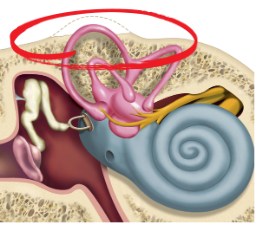
Bow Hunter’s syndrome (BHS) is a rare vascular disorder in which head or neck movement causes dynamic narrowing or occlusion of a vertebral artery, which can compromise blood flow through the posterior circulation. It is also referred to as rotational vertebral artery syndrome or rotational vertebral artery occlusion syndrome (Choi et al., 2026; Elizondo-Ramirez et al., 2024).
BHS is most commonly triggered by head rotation, although flexion and extension can also provoke vascular compromise in some cases. Causes may include cervical spondylosis, osteophytes, or other structural or vascular abnormalities (Choi et al., 2026; Zhu & Wang, 2026). Symptoms can include dizziness, vertigo, imbalance, and nystagmus, making BHS an important vascular differential when symptoms are consistently provoked by cervical movement (Iida et al., 2018; Nomura et al., 2020; Schubert et al., 2021).
History

Dr. Bruce F. Sorenson
Etiology
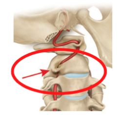
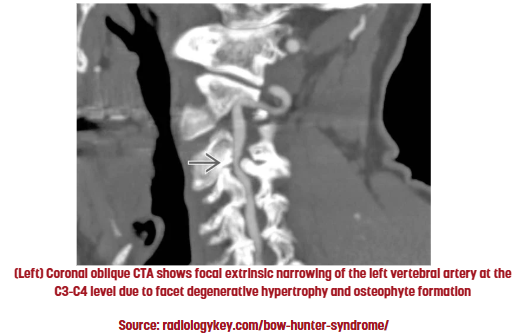
BHS results from dynamic mechanical compromise of a vertebral artery during head or neck movement. Compression may occur in the upper cervical region or at subaxial cervical levels. Degenerative changes, particularly cervical osteophytes and spondylosis, are the most commonly reported structural causes (Elizondo-Ramirez et al., 2024; Zhu & Wang, 2026).
Other reported causes include:
- Cervical disc herniation
- Fibrous bands
- Cervical instability or abnormal vertebral motion
- Congenital bony abnormalities
- Vertebral artery variants, including abnormal origin, course, or hypoplasia
- Rarely, other soft-tissue or musculoskeletal structures that mechanically affect the artery (Choi et al., 2026).
Prevalence
Bow Hunter’s syndrome is rare, and its population prevalence and incidence have not been established. Published evidence consists primarily of case reports and case series rather than population-based epidemiologic studies. A 2024 literature review identified 121 reported patients, with a mean age of 50 years; most were adults, although pediatric and adolescent cases have also been described (Elizondo-Ramirez et al., 2024).
Patient Reported Symptoms
Symptoms are typically triggered by a specific head or neck position, most often rotation, and may improve when the head returns toward neutral (Nomura et al., 2020; Schubert et al., 2021).
Patients may report:
- Vertigo or dizziness
- Imbalance or unsteadiness
- Presyncope or syncope
- Visual changes, including blurred vision, diplopia, or transient vision loss
- Nausea, sometimes with vomiting
- Less commonly, tinnitus or hearing changes
- Other transient neurologic symptoms such as dysarthria, dysphagia, weakness, numbness, or incoordination (Elizondo-Ramirez et al., 2024; Schubert et al., 2021)

Clinical Findings
Key Bedside Clues
- Symptoms are consistently linked to a specific cervical movement and typically improve when the head returns toward neutral.
- Consider a vascular cause when the presentation is atypical for BPPV, particularly when symptoms can be reproduced with cervical rotation in an upright, non-gravity-dependent position (Schubert et al., 2021).
- No bedside test can confirm or exclude Bow Hunter’s syndrome.
Vestibular / Oculomotor Testing
- Rotation-provoked nystagmus may be observed with infrared video goggles.
- Downbeat nystagmus is an important reported clue, but horizontal, torsional, and direction-changing/transitional patterns have also been described (Iida et al., 2018; Nomura et al., 2020; Schubert et al., 2021).
- Baseline oculomotor and vestibular findings may be normal when the provoking position is avoided (Schubert et al., 2021).
Key Take-Home: There is no characteristic nystagmus pattern diagnostic of BHS. The relationship between the eye movements, symptoms, and cervical position is more important than nystagmus direction alone.
Diagnostic Imaging
- Static CTA or MRA may miss BHS when vascular compromise occurs only with movement.
- Dynamic Doppler ultrasound or dynamic CTA may demonstrate position-dependent changes in vertebral artery flow or caliber (Nomura et al., 2020).
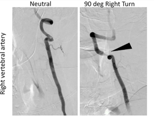
- Dynamic digital subtraction angiography (DSA) is considered the diagnostic gold standard, demonstrating vertebral artery compromise during the provoking movement (Choi et al., 2026).
Treatment
There is no established gold-standard treatment for Bow Hunter’s syndrome. Management is individualized based on the cause and location of vertebral artery compromise, cervical stability, vascular anatomy, and presence of ischemic events (Elizondo-Ramirez et al., 2024; Schulz et al., 2021).
🛑 Conservative Management
May be considered in selected patients and can include:
- Avoiding provocative neck positions
- Cervical collar or temporary immobilization
- Antithrombotic therapy when medically indicated
Conservative management does not correct an underlying structural compression, and some patients with persistent symptoms or recurrent ischemic events ultimately require intervention (Elizondo-Ramirez et al., 2024; Schulz et al., 2021).

🔧 Surgical Management
Surgery is the most commonly reported intervention for symptomatic BHS with a correctable structural cause.
- Decompression removes the structure compressing the vertebral artery.
- Cervical fusion restricts the motion causing vascular compromise and may be considered when instability is present or stabilization is needed.
- The surgical approach depends on the level and mechanism of compression (Elizondo-Ramirez et al., 2024).

🩸Endovascular Treatment
Endovascular treatment is less commonly used and supported by limited evidence. Vertebral artery stenting has been reported in selected cases, particularly when the vascular anatomy and underlying mechanism are suitable for an endovascular approach (Motiei-Langroudi et al., 2017)
💡Key Take-Home:
Treatment is cause-specific and multidisciplinary. For vestibular clinicians, the primary role is to recognize a possible vascular presentation and facilitate appropriate medical referral rather than treating the symptoms as a peripheral positional vestibular disorder.
To Learn More — Check Out These Resources

- Vestibular First Courses:
- Watch related Journal Clubs:
- FREE Patient Resources:
- FREE page:
References
- Choi JW, Qiao Y, Mehta TI, Wilson JN, Torigoe TH, Tsappidi S, Jonathan Zhang Y, Brown SC, Hui FK, Abruzzo T. Safety and efficacy of dynamic catheter-directed cerebral digital subtraction angiography for diagnosis of bowhunter syndrome spectrum disorders: A systematic review of the literature. Interv Neuroradiol. 2026 Aug;32(4):1615-1626. doi: 10.1177/15910199241236820. Epub 2024 Mar 13. PMID: 38477583; PMCID: PMC11577335. https://pmc.ncbi.nlm.nih.gov/articles/PMC11577335/?utm_source
- Devaraja K. Approach to cervicogenic dizziness: a comprehensive review of its aetiopathology and management. Eur Arch Otorhinolaryngol. 2018 Oct;275(10):2421-2433. doi: 10.1007/s00405-018-5088-z. Epub 2018 Aug 9. PMID: 30094486. https://pubmed.ncbi.nlm.nih.gov/30094486/
- Elizondo-Ramirez J, Herrera-Castro JC, Del Rio-Martinez CJ, Torres-Figueroa OJ, Munoz-Cobos A, Ordonez-Solorio LA. Bow Hunter’s syndrome surgical approach and outcome: Two new cases and literature review. Surg Neurol Int. 2024 Nov 8;15:404. doi: 10.25259/SNI_633_2024. PMID: 39640315; PMCID: PMC11618828. https://pubmed.ncbi.nlm.nih.gov/39640315/
- Iida Y, Murata H, Johkura K, Higashida T, Tanaka T, Tateishi K. Bow Hunter’s Syndrome by Nondominant Vertebral Artery Compression: A Case Report, Literature Review, and Significance of Downbeat Nystagmus as the Diagnostic Clue. World Neurosurg. 2018 Mar;111:367-372. doi: 10.1016/j.wneu.2017.12.167. Epub 2018 Jan 5. PMID: 29309982. https://pubmed.ncbi.nlm.nih.gov/29309982/
- Motiei-Langroudi R, Griessenauer CJ, Alturki A, Adeeb N, Thomas AJ, Ogilvy CS. Bow Hunter’s Syndrome from a Tortuous V1 Segment Vertebral Artery Treated with Stent Placement. World Neurosurg. 2017 Feb;98:878.e11-878.e15. doi: 10.1016/j.wneu.2016.11.067. Epub 2016 Nov 22. PMID: 27888081. https://pubmed.ncbi.nlm.nih.gov/27888081/
- Nomura Y, Toi T, Ogawa Y, Oshima T, Saito Y. Transitional nystagmus in a Bow Hunter’s Syndrome case report. BMC Neurol. 2020 Nov 30;20(1):435. doi: 10.1186/s12883-020-02009-3. PMID: 33256636; PMCID: PMC7706255. https://pubmed.ncbi.nlm.nih.gov/33256636/
- Schubert MC, Carter N, Lo SL. Case Report: Bow Hunter Syndrome-One Reason to Add Non-gravity Dependent Positional Nystagmus Testing to Your Clinical Neuro-Otologic Exam. Front Neurol. 2021 Dec 20;12:814998. doi: 10.3389/fneur.2021.814998. PMID: 34987472; PMCID: PMC8720997. https://pubmed.ncbi.nlm.nih.gov/34987472/
- Schulz R, Donoso R, Weissman K. Rotational vertebral artery occlusion (“bow hunter syndrome”). Eur Spine J. 2021 Jun;30(6):1440-1450. doi: 10.1007/s00586-020-06680-5. Epub 2021 Jan 2. PMID: 33389200. https://pubmed.ncbi.nlm.nih.gov/33389200/
- Zhu F, Wang H. Bow hunter’s syndrome secondary to lateral and posterior spondylosis: illustrative case. J Neurosurg Case Lessons. 2026 Mar 9;11(10):CASE25718. doi: 10.3171/CASE25718. PMID: 41802288; PMCID: PMC12970371. https://pmc.ncbi.nlm.nih.gov/articles/PMC12970371/?utm_source