
Half Dix-Hallpike Test
Fundamentals
The half Dix-Hallpike is a supplemental positional test classically described for suspected posterior cupulolithiasis BPPV. The standard Dix-Hallpike test remains the primary test for posterior canal BPPV, while the half Dix-Hallpike may provide additional information when findings are atypical or inconclusive (Wang et al., 2022; Kim et al., 2026).
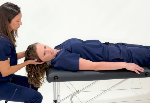
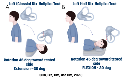
In the standard half Dix-Hallpike, the head is turned 45° toward the side being tested and positioned approximately 30° above horizontal. This orients the posterior canal cupula approximately perpendicular to gravity, allowing a weighted cupula to be maximally deflected (von Brevern et al., 2015; Kim et al., 2026). Persistent upbeat-torsional nystagmus, with torsion toward the affected ear and lasting longer than 1 minute, supports posterior canal cupulolithiasis (von Brevern et al., 2015; Wu et al., 2024).
An augmented half Dix-Hallpike has also been described as an emerging test for suspected short-arm posterior canal canalithiasis. The patient is first held in the conventional Dix-Hallpike position and then raised into the half Dix-Hallpike position. This sequence is proposed to move short-arm otoconia toward the cupula before positioning the cupula perpendicular to gravity (Kim et al., 2026). It may be useful when the standard Dix-Hallpike produces little or no nystagmus despite findings suggestive of posterior canal BPPV (Kim et al., 2026).
The overall diagnostic sensitivity of the half Dix-Hallpike has not been established. False-negative responses can occur, meaning posterior canal cupulolithiasis may still be present even when characteristic nystagmus is not elicited; findings should therefore be interpreted with other positional tests (Wang et al., 2022; Kim et al., 2026).
History

The half Dix-Hallpike can be traced to Epley’s work in 2001. The test later gained greater clinical recognition when the Bárány Society’s 2015 BPPV diagnostic criteria included the half Dix-Hallpike in the assessment of posterior canal cupulolithiasis.
More recent studies have further evaluated its diagnostic value and limitations in atypical posterior canal BPPV (Wang et al., 2022; Wu et al., 2024; Kim et al., 2026).
Instructions
The goal is to help identify posterior canal cupulolithiasis by positioning the posterior canal cupula so gravity can more effectively provoke its characteristic nystagmus.
Right Ear
- Explain the procedure to the patient and obtain consent.
- Position the patient in long-sitting on the exam table and instruct the patient to keep their eyes open throughout the test.
- Have the patient turn their head 45 degrees to the right.
- Keep the head to the right, support the head and guide the patient into a supine position with the head flexed about 30° with the patient’s shoulders resting on a surface (table, bed, or pillow).
- Observe the patient’s eyes for nystagmus and ask about vertigo symptoms.
- Hold this position at least 30 to 60 sec.
- If not moving into treatment from the testing position, assist the patient back to sitting, maintaining head rotation to the right.
- Monitor for vertigo and nystagmus during return to sitting and continue to guard the patient.
Left Ear
- Explain the procedure to the patient and obtain consent.
- Position the patient in long-sitting on the exam table and instruct the patient to keep their eyes open throughout the test.
- Have the patient turn their head 45 degrees to the left.
- Keep the head to the left, support the head and guide the patient into a supine position with the head flexed about 30° with the patient’s shoulders resting on a surface (table, bed, or pillow).
- Observe the patient’s eyes for nystagmus and ask about vertigo symptoms.
- Hold this position for 30 to 60 seconds.
- If not moving into treatment from the testing position, assist the patient back to sitting, maintaining head rotation to the left.
- Monitor for vertigo and nystagmus during return to sitting and continue to guard the patient.
Test Interpretation
Related Pathology
- Posterior Cupulolithiasis
- Short-arm posterior canal canalithiasis: an emerging application of the augmented Half Dix-Hallpike, particularly when the standard Dix-Hallpike is negative or atypical (Kim et al., 2026).
The half Dix-Hallpike test is best done with infrared video goggles due the risk of otherwise missing about 2/3 of abnormal eye movements when testing in room light or with traditional thickened lens Frenzels. See what you’re missing – access a free virtual demo (on-demand or live with a clinician) today.
Sources
- Helminski JO. Atypical PC-BPPV—cupulolithiasis and short-arm canalithiasis: a retrospective observational study. J Neurol Phys Ther. 2025;49(1):51-61. https://www.ovid.com/jnls/jnpt/fulltext/10.1097/npt.0000000000000494~atypical-pc-bppv-cupulolithiasis-and-short-arm-canalithiasis
- Kim HJ, Gil YE, Kim JS. Cupulolithiatic BPPV involving both posterior semicircular canals: implications for set-point adaptation. J Neurol. 2024 Nov;271(11):7325-7329. doi: 10.1007/s00415-024-12683-9. Epub 2024 Sep 14. PMID: 39277569.
-
Kim H-J, Kim J-S, Büki B. New developments in the diagnosis of benign paroxysmal positional vertigo. Journal of Vestibular Research. 2026;0(0). doi:10.1177/09574271261454144
- Wang W, Yan S, Zhang S, et al. Clinical application of different vertical position tests for posterior canal-benign paroxysmal positional vertigo-cupulolithiasis. Front Neurol. 2022;13:930542. https://www.frontiersin.org/journals/neurology/articles/10.3389/fneur.2022.930542/full
- von Brevern M, Bertholon P, Brandt T, et al. Benign paroxysmal positional vertigo: diagnostic criteria: consensus document of the Committee for the Classification of Vestibular Disorders of the Bárány Society. J Vestib Res. 2015;25(3-4):105-117. doi:10.3233/VES-150553. https://journals.sagepub.com/doi/pdf/10.3233/VES-150553
- Wu J, Zou Y, Xu W, et al. Retrospective analysis of nystagmus characteristics and clinical applications of positional testing in patients with cupulolithiasis of the posterior semicircular canal in benign paroxysmal positional vertigo. Front Neurol. 2024;15:1413929. https://pmc.ncbi.nlm.nih.gov/articles/PMC11266046/