
Quick Liberatory Rotation
Description
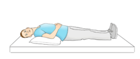
Quick Liberatory Rotation (QLR) is a rapid, clinician-performed maneuver used for long arm posterior canalithiasis BPPV and apogeotropic posterior canal canalithiasis BPPV. It combines features of repositioning and liberatory maneuvers: the movement pathway is similar to posterior canal repositioning techniques, while the speed of the rotation is more characteristic of a liberatory maneuver (Califano et al., 2003; Califano et al., 2014).
QLR is not usually listed as a standard treatment for posterior cupulolithiasis, although modified or vibration-assisted approaches involving QLRM have appeared in limited atypical case reports.
Effectiveness
For typical long arm posterior canalithiasis BPPV, Quick Liberatory Rotation has been reported to resolve BPPV in about 79–80% of patients after one treatment, with the original study reporting up to 98% resolution within three treatment cycles (Califano et al., 2003; Califano et al., 2014).
For apogeotropic posterior canal BPPV, the evidence is more limited. One case series reported direct resolution in 8 of 23 cases, while 14 of 23 cases converted into typical posterior canal BPPV and were then treated with QLR as a second step (Califano et al., 2014).
History
Quick Liberatory Rotation was developed by Luigi Califano and colleagues at “G. Rummo” Hospital in Benevento, Italy, and was first used clinically in July 2001. Published in 2003, QLR reflected an effort to refine posterior canal BPPV treatment by pairing the anatomical logic of canalith repositioning with the rapid movement strategy of a liberatory maneuver. Its name highlights the feature that made it distinct: a fast rotation intended to move posterior canal debris efficiently while allowing the clinician to observe treatment-related nystagmus (Califano et al., 2003).

Dr. Luigi Califano
Instructions
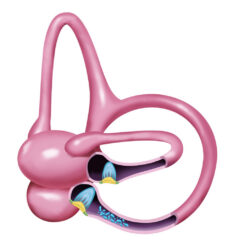
The purpose is to move loose otoconia out of the posterior semicircular canal and back toward the utricle, where they are less likely to trigger positional vertigo.
Right
- Begin with the patient sitting.
- Perform a right Dix-Hallpike test to place the patient into the affected-side Dix-Hallpike position.
- Wait until the nystagmus stops. Then wait 2 additional minutes.
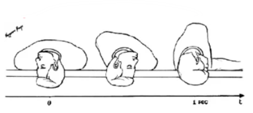
- Quickly rotate the patient from the right Dix-Hallpike position to the left side-lying position.
- The rotation should be fast: less than 1 second, with the head moving about 180° in the horizontal plane.
- End with the patient lying on the left side, with the head angled about 45° downward.
- In this final position, observe for secondary nystagmus similar in direction to the diagnostic Dix-Hallpike nystagmus.
- Hold this final position for 2 minutes after secondary nystagmus disappears. If no secondary nystagmus is seen, hold the position for 4 minutes.
- Return the patient to sitting.
Left
- Begin with the patient sitting.
- Perform a left Dix-Hallpike test to place the patient into the affected-side Dix-Hallpike position.
- Wait until the nystagmus stops. Then wait 2 additional minutes.
- Quickly rotate the patient from the left Dix-Hallpike position to the right side-lying position.
- The rotation should be fast: less than 1 second, with the head moving about 180° in the horizontal plane.
- End with the patient lying on the right side, with the head angled about 45° downward.
- In this final position, observe for secondary nystagmus similar in direction to the diagnostic Dix-Hallpike nystagmus.
- Hold this final position for 2 minutes after secondary nystagmus disappears. If no secondary nystagmus is seen, hold the position for 4 minutes.
- Return the patient to sitting.
Related Pathology
Sources
- Califano L, Capparuccia PGG, Di Maria D, Melillo MG, Villari D. Treatment of benign paroxysmal positional vertigo of posterior semicircular canal by “Quick Liberatory Rotation Manoeuvre.” Acta Otorhinolaryngol Ital. 2003;23(3):161–167. https://old.actaitalica.it/issues/2003/3_03/02.%20CALIFANO.pdf
- Califano L, Salafia F, Mazzone S, Melillo MG, Califano M. Double-Blind Randomized Trial on the Efficacy in a Short-Time Follow-Up of the “Quick Liberatory Rotation” Maneuver in Treating Posterior Canal BPPV. Journal of Otolaryngology Advances. 2014;1(1):1–11. doi:10.14302/issn.2379-8572.joa-14-418. https://openaccesspub.org/otolaryngology-advances/article/double-blind-randomized-trial-on-the-efficacy-in-a-short-time-follow-up-of-the-quick-liberatory-rotation-maneuver-in-treating-posterior-cana-118
- Califano L, Salafia F, Mazzone S, Melillo MG, Califano M. Anterior canal BPPV and apogeotropic posterior canal BPPV: two rare forms of vertical canalolithiasis. Acta Otorhinolaryngol Ital. 2014;34(3):189-197. https://old.actaitalica.it/issues/2014/3-2014/06-califano.pdf?utm_source
- Vats AK, Castellucci A, Kothari S, Vats S, Patil M. Direction-Reversing Positional Nystagmus in Typical Posterior Semicircular Canalolithiasis with Ipsicanal Transformation via Non-ampullary Arm Canalolithiasis to Posterior Cupulolithiasis. Ann Indian Acad Neurol. 2025;28(3):478-479. doi:10.4103/aian.aian_842_24 https://www.researchgate.net/publication/390928845_Direction-Reversing_Positional_Nystagmus_in_Typical_Posterior_Semicircular_Canalolithiasis_with_Ipsicanal_Transformation_via_Non-ampullary_Arm_Canalolithiasis_to_Posterior_Cupulolithiasis