
Minimum Stimulus Strategy: Upright BPPV Testing
Fundamentals
The Minimum Stimulus Strategy (MSS) is a nystagmus-guided approach used to identify the involved canal, side, and BPPV pattern while limiting provocative positioning.
Its Upright BPPV Protocol is performed in sitting. Pitch is forward-and-backward head movement, while roll is tilting one ear toward the shoulder. The vertical canals form diagonal pairs: RALP includes the right anterior and left posterior canals, while LARP includes the left anterior and right posterior canals. For posterior-canal testing, turning the head 45° left before pitching tests the left posterior canal through uRALP; turning right tests the right posterior canal through uLARP (Libonati et al., 2022).
In confirmed BPPV cohorts, the upright protocol agreed with the complete assessment in 95.5% of lateral-canal cases. For posterior-canal BPPV, the Head Pitch Test identified 75.2%, increasing to 87.2% with uRALP/uLARP testing. The combined protocol identified 100% of patients tested less than seven days after symptom onset, compared with 68.9% tested later (Martellucci et al., 2020; Martellucci et al., 2022).
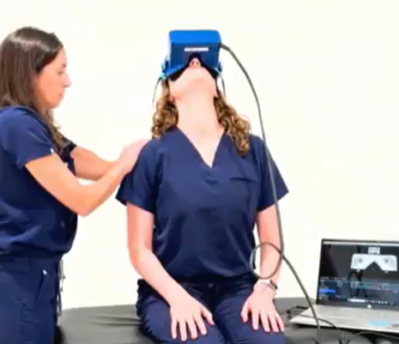
Upright testing may reduce supine or head-hanging positions and may be especially valuable for highly symptomatic patients or those who poorly tolerate these positions. It can reduce discomfort and autonomic symptoms associated with provocative supine testing. Nystagmus should be monitored continuously with infrared video goggles, and conventional positional testing should follow when findings are absent, unclear, or atypical (Martellucci et al., 2020; Libonati et al., 2022).
History
Dr. Asprella Libonati

Dr. Salvatore Martellucci
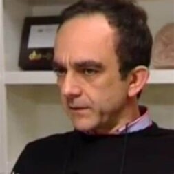
The Minimum Stimulus Strategy began with Dr. Asprella Libonati’s in 2003 and was formally described in 2005 as a way to diagnose and treat BPPV while provoking fewer vertigo episodes (Asprella Libonati et al., 2003; Asprella Libonati, 2005).
In 2020, the Upright Head Roll Test was incorporated into a complete upright protocol for horizontal or lateral-canal BPPV. In 2022, the strategy expanded to include uRALP and uLARP testing for the vertical canals, followed by additional validation studies in 2022 and 2024 (Malara et al., 2020; Martellucci et al., 2020; Libonati et al., 2022; Martellucci et al., 2022; Han et al., 2024).
Instructions
The purpose of upright BPPV testing is to use seated nystagmus findings to guide the next diagnostic or treatment step. It provides a less provocative option when conventional positional testing is poorly tolerated.
Preparation
- Seat the patient upright with the head facing forward.
- Use infrared video goggles to remove visual fixation and continuously monitor nystagmus.
- Screen cervical mobility before testing. When neck motion is limited, the trunk may be tilted approximately 30° in the testing plane to achieve a similar head position relative to gravity (Martellucci et al., 2020; Libonati et al., 2022).
Upright Testing Protocol
Neutral Sitting
Observe the patient with the head aligned with the trunk and the eyes looking forward. Note any persistent horizontal nystagmus before moving the head.
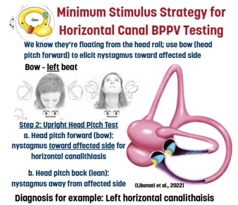
When horizontal nystagmus is present, perform the Upright Head Pitch Test to determine whether its direction changes with head angle. This direction-changing pattern supports pseudo-spontaneous nystagmus associated with horizontal or lateral-canal BPPV; neutral observation alone cannot confirm or lateralize BPPV (Libonati et al., 2022; Martellucci et al., 2020).
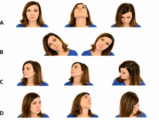
A. Upright Head Pitch Test
- Slowly flex the head approximately 60° forward.
- Hold until nystagmus appears, usually within 30 seconds.
- Move through neutral and extend the head approximately 30° backward.
- Hold and observe again.
The test may produce direction-changing horizontal nystagmus with horizontal canal BPPV or vertical nystagmus with variable torsional components when a vertical canal is involved (Libonati et al., 2022; Martellucci et al., 2020).
The next test depends on the findings:
- Horizontal nystagmus: proceed to the Upright Head Roll Test.
- Vertical nystagmus with absent or unclear torsion: proceed to uRALP and uLARP testing.
B. Upright Head Roll Test
Use when horizontal nystagmus suggests horizontal canal involvement.
- Slowly tilt the head approximately 30° toward one shoulder without rotating it.
- Hold until nystagmus appears, usually within 30 seconds.
- Return to neutral and hold for approximately 30 seconds.
- Repeat toward the opposite shoulder.
- Record the direction of nystagmus in each position.
The Upright Head Roll Test follows a positive horizontal Head Pitch Test within the published lateral-canal protocol (Malara et al., 2020; Martellucci et al., 2020).
C. Upright RALP Test: Left Posterior Canal
Use when left posterior-canal BPPV is suspected or the Head Pitch Test does not show clear torsion.
- Rotate the head 45° to the left.
- Maintain the rotation while slowly flexing the head 60° forward.
- Move through neutral and extend the head 30° backward.
- Observe for nystagmus in both positions.
The RALP plane contains the right anterior and left posterior canals and approximately aligns head movement with the left posterior canal (Libonati et al., 2022).
D. Upright LARP Test: Right Posterior Canal
Use when right posterior-canal BPPV is suspected or the Head Pitch Test does not show clear torsion.
- Rotate the head 45° to the right.
- Maintain the rotation while slowly flexing the head 60° forward.
- Move through neutral and extend the head 30° backward.
- Observe for nystagmus in both positions.
The LARP plane contains the left anterior and right posterior canals and approximately aligns head movement with the right posterior canal (Libonati et al., 2022).
Test Interpretation
Posterior-Canal BPPV During the Head Pitch Test
| Head position | Expected nystagmus |
|---|---|
| Forward flexion | Downbeat with torsion toward the unaffected ear |
| Backward extension | Upbeat with torsion toward the affected ear |
Torsion is described by the direction of the fast phase at the upper poles of the eyes (Libonati et al., 2022).
Upright RALP and LARP Findings
| Suspected canal | Forward flexion | Extension |
|---|---|---|
| Left posterior: uRALP |
Downbeat with right torsion | Upbeat with left torsion |
| Right posterior: uLARP |
Downbeat with left torsion | Upbeat with right torsion |
These tests may make torsion easier to detect by aligning the movement more closely with the suspected posterior canal (Libonati et al., 2022).
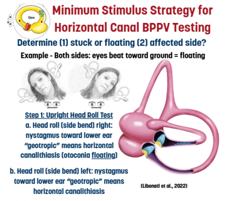
Upright Head Roll Findings
| Nystagmus pattern | Interpretation |
|---|---|
|
Beats toward the |
Geotropic horizontal canalithiasis |
| Beats toward the upper ear during both tilts |
Apogeotropic horizontal cupulolithiasis |
The Head Pitch and Upright Head Roll findings should be interpreted together to determine the likely lateral-canal variant and side.

Atypical or Inconclusive Findings
Downbeat nystagmus in both forward and backward positions, particularly when torsion is absent or difficult to detect, may occur with anterior-canal BPPV, apogeotropic posterior-canal BPPV or short arm posterior canal BPPV. These conditions may remain difficult to distinguish using positional nystagmus alone (Libonati et al., 2022).
Positional downbeat nystagmus can also have a central cause. Further neurologic and vestibular evaluation is warranted when findings do not follow an expected canal plane, are persistent or otherwise atypical, or occur with additional neurologic or oculomotor abnormalities (Macdonald et al., 2017).
Proceed with conventional positional testing when:
- Upright testing does not evoke nystagmus.
- Torsion or the involved side remains unclear.
- The findings do not match an expected canal-specific pattern.
- A central or non-BPPV cause remains possible.
After identifying a suspected posterior-canal side with upright testing, the MSS authors recommend confirming the diagnosis with an ipsilateral Dix–Hallpike or side-lying diagnostic test before treatment. For suspected lateral-canal BPPV, proceed to supine positioning and the supine roll test when the upright findings do not provide a reliable diagnosis (Libonati et al., 2022; Martellucci et al., 2020).
Watch Related Journal Club
Minimum Stimulus BPPV Testing with Dr. Castellucii and Dr. Martellucci
Related Pathology
Minimum Stimulus Testing is best done with infrared video goggles due the risk of otherwise missing about 2/3 of abnormal eye movements when testing in room light or with traditional thickened lens Frenzels. See what you’re missing – access a free virtual demo (on-demand or live with a clinician) today.
Sources
- Asprella Libonati G, Gagliardi G, Cifarelli D, Larotonda G. “Step by step” treatment of lateral semicircular canal canalolithiasis under videonystagmoscopic examination. Acta Otorhinolaryngol Ital. 2003;23(1):10-15. PubMed | Full text
- Asprella Libonati G. Diagnostic and treatment strategy of lateral semicircular canal canalolithiasis. Acta Otorhinolaryngol Ital. 2005;25(5):277-283. PubMed | Full text
- Han JS, Lee DH, Park SN, et al. Evaluation of the reliability and validity of the upright head roll test for lateral semicircular canal benign paroxysmal positional vertigo. J Vestib Res. 2024;34(2-3):103-112. doi:10.3233/VES-230127. PubMed | Journal article
- Libonati GA, Martellucci S, Castellucci A, Malara P. Minimum Stimulus Strategy: a step-by-step diagnostic approach to BPPV. J Neurol Sci. 2022;434:120158. doi:10.1016/j.jns.2022.120158. PubMed | Journal abstract
- Malara P, Castellucci A, Martellucci S. Upright head roll test: a new contribution for the diagnosis of lateral semicircular canal benign paroxysmal positional vertigo. Audiol Res. 2020;10(1):236. doi:10.4081/audiores.2020.236. Full text
- Martellucci S, Malara P, Castellucci A, et al. Upright BPPV Protocol: feasibility of a new diagnostic paradigm for lateral semicircular canal benign paroxysmal positional vertigo compared to standard diagnostic maneuvers. Front Neurol. 2020;11:578305. doi:10.3389/fneur.2020.578305. Full text
- Martellucci S, Castellucci A, Malara P, et al. Is it possible to diagnose posterior semicircular canal BPPV from the sitting position? The role of the Head Pitch Test and the upright tests along the RALP and LARP planes. Am J Otolaryngol. 2022;43(4):103474. doi:10.1016/j.amjoto.2022.103474. PubMed | Journal abstract